
The exact cause of bipolar disorder is unknown, but it is thought to be a combination of genetic and environmental factors. Brain imaging studies have shown that people with bipolar disorder have differences in the structure and function of certain brain areas, including the frontal lobe, temporal lobe, and limbic system.
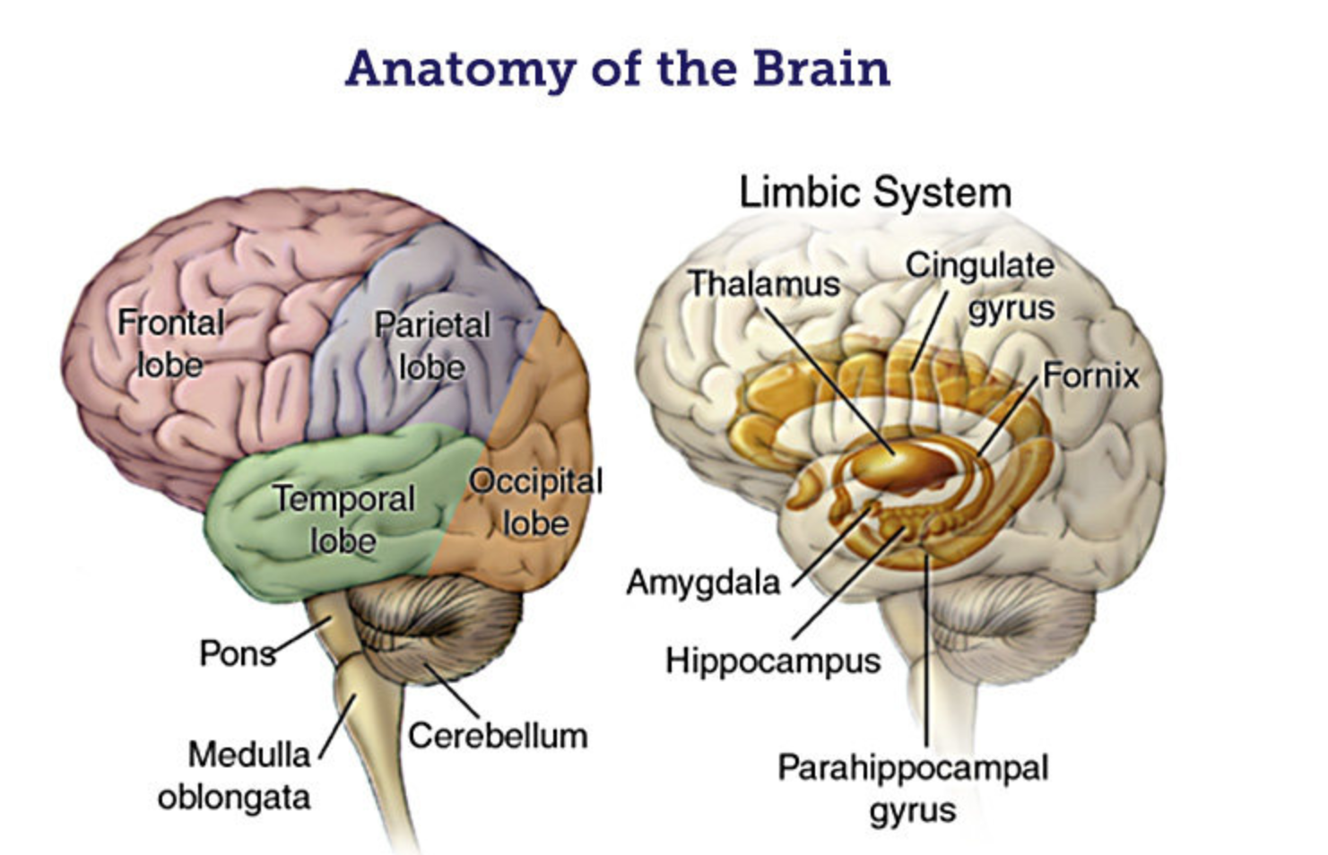
- The frontal lobe is involved in decision-making, planning, and impulse control. People with bipolar disorder may have difficulty making decisions, planning ahead, and controlling their impulses.
- The temporal lobe is involved in memory, emotion, and social behavior. People with bipolar disorder may have problems with memory, mood regulation, and social interactions.
- The limbic system is involved in emotion, motivation, and behavior. People with bipolar disorder may have intense emotions, be easily motivated, and engage in risky behaviors.
A review of articles (a meta-analysis) published in the research journal JAMA Psychiatry looked at regional brain activity in bipolar individuals compared with people without bipolar disorder when people performed three types of tasks.
- emotion processing tasks (for example, evaluating pictures of people experiencing different emotions).
- reward processing tasks (for example, using information about possible rewards to make decisions about what to do).
- working memory tasks (for example, tasks that rely on holding multiple pieces of information in immediate memory).
Individuals with bipolar disorder (BD) have different brain activity patterns than those without BD during these three types of tasks.
When processing emotions, individuals with BD show more activity in the amygdala and less connectivity between the ventrolateral prefrontal cortex (PFC), anterior cingulate cortex (ACC), orbitofrontal cortex (OFC), and limbic areas. Increased activity in the amygdala, without the moderating influence of the cortex, may be associated with greater emotionality.

When processing rewards, individuals with BD show more activity in the PFC, ACC, and striatum.
When working on memory tasks, individuals with BD show different activity patterns in the frontal cortex.
These findings have not been consistent across all studies, but they are found in most studies.
Mood Dependent or Mood Independent Changes
Some of the differences in brain activity were seen in bipolar people whether or not they were acutely depressed or manic and some of the differences were seen only in those who were acutely depressed or manic.
Findings that occurred in all moods in bipolar individuals are called trait changes (they are due to bipolar disorder itself rather than due to being in a temporary state of depression or mania). Trait changes seen in these studies almost always reflected changes in limbic activity.
It’s possible that the changes in limbic system activity in people with bipolar during periods of normal mood may play a role in the extreme mood shifts seen in people with bipolar.
Emotion Processing Changes
These are the findings in bipolar patients that are not mood dependent:
- During emotion processing people with bipolar had a mood independent increase in activity in the hippocampus, parts of the temporal cortex, and amygdala.
- Also, there was reduced activity during emotion processing in the right inferior frontal gyrus (IFG) that was not affected by mood changes.
When there are changes in mood, altered frontal cortex activity may be involved in the persistence of moods.
Mood dependent alterations in activation of brain regions when emotion processing were:
- When in a normal mood state, bipolar individuals had increased activity in the left parahippocampal gyrus and reduced activity in the left IFG.
- During mania, the authors of the study found increased activity in the left thalamus and reduced activity in the right IFG.
- When depressed, people with bipolar had increased activity in the left parietal lobe.
Reward Processing Changes
The only consistent change in brain activity during reward processing tasks across all mood states was increased activity of the left orbitofrontal cortex OFC.
In a normal mood, people with bipolar had increased activity in the left parahippocampal gyrus, ACC, MFG, and right temporal gyrus.
Individuals who were depressed had increased activity in the left inferior frontal and in the right superior temporal gyrus.
There were not enough studies of manic people to see differences.
Working Memory Changes
Across all moods, people with bipolar had increased activity in the left subgenual ACC and ventromedial PFC. Manic individuals had increased activity in the left ACC and reduced activity in the left IFG during working memory. Depressed people with bipolar had increased activity in the left PFC and ACC, and reduced activity in the right parietal lobe and left cerebellum.
References
Mesbah R, Koenders MA, van der Wee NJA, Giltay EJ, van Hemert AM, de Leeuw M. Association Between the Fronto-Limbic Network and Cognitive and Emotional Functioning in Individuals With Bipolar Disorder: A Systematic Review and Meta-analysis. JAMA Psychiatry. Published online March 29, 2023. doi:10.1001/jamapsychiatry.2023.0131